Gastrointestinal (GI) tissue biopsies present vital diagnostic info for all kinds of situations similar to neoplastic illnesses (colorectal, small bowel and abdomen cancers) and non-neoplastic illnesses (inflammatory problems, an infection, celiac illness). Endoscopic biopsies acquire small tissue samples that require useful resource intensive processing to allow histopathological evaluation. Unfortunately, the sparsely collected biopsy samples might fail to seize the pathologic situation as a result of choice of biopsy websites depends on macroscopic superficial tissue options and clinician judgement. Here, we current the primary all-optical non-contact label-free non-interferometric photoacoustic microscopy system succesful of performing “virtual biopsies”.
A modular photoacoustic remote sensing (PARS™) structure is used facilitating imaging of unprocessed tissues offering info just like standard histopathological staining strategies. Prospectively this might permit gastroenterologists to evaluate subcellular tissue morphology in situ when deciding on biopsy location. Tested on preserved unstained human and freshly resected murine tissues, the introduced PARS microscope quickly retrieves photos of comparable space to present biopsies, whereas sustaining comparable high quality to the present customary for histopathological evaluation.
Additionally, outcomes present the primary label free evaluation of subsurface mobile morphology in FFPE GI tissue blocks. Clinically related options are recovered together with mobile particulars similar to lamina propria inside colon tissue and cell nuclear construction in resected easy muscle. Constructed with a modular structure, this method facilitates the longer term growth of compact imaging heads. The modular PARS system overcomes many of the challenges with imaging unstained thick tissue in situ, representing a big milestone within the growth of a scientific microscope offering virtual biopsy capabilities.
Plant cell partitions consist of completely different polysaccharides and structural proteins, which kind a inflexible layer situated outdoors of the plasma membrane. The wall can be a really dynamic cell composite, which is characterised by complicated polysaccharide interactions and varied modifications throughout cell growth. The visualization of cell wall parts in situ may be very difficult because of the small measurement of cell wall composites (nanometer scale), giant variety of the wall polysaccharides and their complicated interactions.
This protocol describes immunogold labeling of completely different cell wall epitopes for high-resolution transmission electron microscopy (TEM). It supplies an in depth process for assortment and preparation of plant materials, ultra-thin sectioning, specimen labeling and contrasting. An immunolabeling process workflow was optimized to acquire excessive effectivity of carbohydrates labeling for high-resolution TEM. This methodology was utilized to check plant cell wall traits in varied plant tissues however may be utilized for different cell parts in plant and animal tissues.
Fluorescence Microscopy Assay to Measure HIV-1 Capsid Uncoating Kinetics in vitro
The stability of the HIV-1 capsid and the spatiotemporal management of its disassembly, a course of referred to as uncoating, should be finely tuned for an infection to proceed. Biochemical strategies for measuring capsid lattice disassembly in bulk are unable to resolve intermediates within the uncoating response. We have developed a single-particle fluorescence microscopy methodology to observe the real-time uncoating kinetics of genuine HIV capsids in vitro.
The assay makes use of immobilized viral particles which might be permeabilized with the a pore-former protein, and is designed to (1) detect the primary defect of the capsid by the discharge of an answer section marker (GFP) and (2) visualize the disassembly of the capsid over time by “portray” the capsid lattice with labeled cyclophilin A (CypA), a protein that binds weakly to the surface of the capsid. This novel assay permits the research of dynamic interactions of molecules with lots of of particular person capsids in addition to to find out their impact on viral capsid stability, which supplies a strong device for dissecting uncoating mechanisms and for the event of capsid-binding medicine.

Workflow in the direction of automated segmentation of agglomerated, non-spherical particles from electron microscopy photos using synthetic neural networks
We current a workflow for acquiring absolutely skilled synthetic neural networks that may carry out automated particle segmentations of agglomerated, non-spherical nanoparticles from scanning electron microscopy photos “from scratch”, with out the necessity for big coaching information units of manually annotated photos. The entire course of solely requires about 15 min of hands-on time by a consumer and may usually be completed inside lower than 12 h when coaching on a single graphics card (GPU). After coaching, SEM picture evaluation could be carried out by the synthetic neural community inside seconds.
This is achieved by using unsupervised studying for many of the coaching dataset era, making heavy use of generative adversarial networks and particularly unpaired image-to-image translation by way of cycle-consistent adversarial networks. We evaluate the segmentation masks obtained with our prompt workflow qualitatively and quantitatively to state-of-the-art strategies using varied metrics. Finally, we used the segmentation masks for routinely extracting particle measurement distributions from the SEM photos of TiO2 particles, which have been in glorious settlement with particle measurement distributions obtained manually however may very well be obtained in a fraction of the time.
) Lugols Iodine Solution (for Microscopy) |
|
RRSP84-F |
Atom Scientific |
2.5L |
EUR 19.31 |
 for microscopy) Hematoxylin solution (Harris) for microscopy |
|
40362 |
Sisco Laboratories |
125 ml |
EUR 4.45 |
|
|
|
Description: Part A |
 for microscopy) Hematoxylin solution (Ehrlich) for microscopy |
|
78631 |
Sisco Laboratories |
125 ml |
EUR 3.35 |
|
|
|
Description: Part A |
 for microscopy) Hematoxylin solution (Mayer`s) for microscopy |
|
48441 |
Sisco Laboratories |
100 ml |
EUR 1.85 |
|
|
|
Description: Part A |
 for microscopy) Hematoxylin solution (Delafield`s) for microscopy |
|
39758 |
Sisco Laboratories |
125 ml |
EUR 3.35 |
|
|
|
Description: Part A |
 Leishman's Stain Solution |
|
S018S-250ML |
EWC Diagnostics |
1 unit |
EUR 4.74 |
|
Description: Leishman's Stain Solution |
 Gentian violet for microscopy |
|
GRM6354-100G |
EWC Diagnostics |
1 unit |
EUR 12.6 |
|
Description: Gentian violet for microscopy |
Gentian violet for microscopy |
|
GRM6354-1KG |
EWC Diagnostics |
1 unit |
EUR 72.01 |
|
Description: Gentian violet for microscopy |
Gentian violet for microscopy |
|
GRM6354-25G |
EWC Diagnostics |
1 unit |
EUR 6.93 |
|
Description: Gentian violet for microscopy |
 ReadiUseâ„¢ microscope mounting solution |
|
20009 |
AAT Bioquest |
50 mL |
EUR 88 |
|
Description: The mounting medium is the solution in which the specimen is embedded, generally under a cover glass. |
ReadiUseâ„¢ microscope mounting solution |
|
20009-50mL |
AAT Bioquest |
50 mL |
EUR 86 |
|
|
|
Description: The mounting medium is the solution in which the specimen is embedded, generally under a cover glass. |
, 95%) Sudan IV for microscopy (C.I. No. 26105), 95% |
|
57335 |
Sisco Laboratories |
25 Gms |
EUR 1.64 |
|
|
|
Description: Part A |
 Cedarwood Oil Natural for microscopy |
|
31018 |
Sisco Laboratories |
30 ml |
EUR 4.99 |
|
|
|
Description: Part A |
 Canada Balsam Natural for microscopy |
|
74616 |
Sisco Laboratories |
100 ml |
EUR 2.85 |
|
|
|
Description: Part B |
, 90%) Thioflavin T for microscopy (C.I. No. 49005), 90% |
|
75493 |
Sisco Laboratories |
5 Gms |
EUR 10.26 |
|
|
|
Description: Part A |
 Immersion Oil Synthetic for microscopy |
|
73017 |
Sisco Laboratories |
30 ml |
EUR 2.19 |
|
|
|
Description: Part A |
 for microscopy) Ethyl Violet (C.I. No. 42600) for microscopy |
|
50371 |
Sisco Laboratories |
25 Gms |
EUR 6.02 |
|
|
|
Description: Part A |
 Fluorescence Microscopy Aid |
|
4400 |
Virostat |
3 ml |
EUR 216.6 |
|
Description: This is Counter - Stain/Blocking Diluent with optimal formula for preparaiton of final dilutions of FITC conjugate prior to use in FA procedures. |
Fluorescence Microscopy Aid |
|
MBS320455-01mg |
MyBiosource |
0.1mg |
EUR 230 |
Fluorescence Microscopy Aid |
|
MBS320455-5x01mg |
MyBiosource |
5x0.1mg |
EUR 945 |
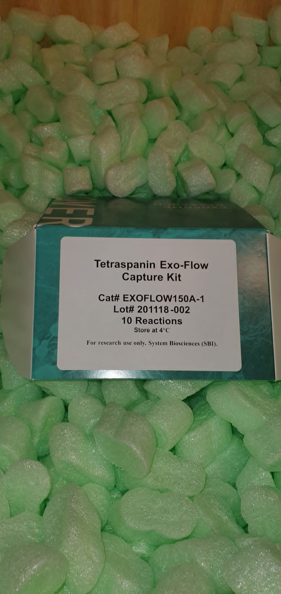
) Exosome electron microscopy imaging (EM) |
|
S109 |
101Bio |
- |
EUR 600 |
, Red Fluorescence) EZClick? Stearoylated Protein Assay Kit (FACS/Microscopy), Red Fluorescence |
|
K418-100 |
Biovision |
each |
EUR 588 |
, Green Fluorescence) EZClick? Stearoylated Protein Assay Kit (FACS/Microscopy), Green Fluorescence |
|
K453-100 |
Biovision |
each |
EUR 588 |
, Red Fluorescence) EZClick? Palmitoylated Protein Assay Kit (FACS/Microscopy), Red Fluorescence |
|
K416-100 |
Biovision |
each |
EUR 588 |
, Red Fluorescence) EZClick? Global RNA Synthesis Assay Kit (FACS/Microscopy), Red Fluorescence |
|
K462-100 |
Biovision |
each |
EUR 639.6 |
EZClick? Global RNA Synthesis Assay Kit (FACS/Microscopy), Red Fluorescence |
|
K718-100 |
Biovision |
each |
EUR 639.6 |
 Stop Solution for TMB Microwell Substrate |
|
6343 |
Immunochemistry |
1000 mL |
EUR 146.5 |
Stop Solution for TMB Microwell Substrate |
|
MBS258088-1L |
MyBiosource |
1L |
EUR 260 |
Stop Solution for TMB Microwell Substrate |
|
MBS258088-5x1L |
MyBiosource |
5x1L |
EUR 1035 |
, Green Fluorescence) EZClick? Myristoylated Protein Assay Kit (FACS/Microscopy), Green Fluorescence |
|
K497-100 |
Biovision |
each |
EUR 588 |
, Green Fluorescence) EZClick? Palmitoylated Protein Assay Kit (FACS/Microscopy), Green Fluorescence |
|
K452-100 |
Biovision |
each |
EUR 588 |
, Red Fluorescence) EZClick? Global Protein Synthesis Assay Kit (FACS/Microscopy), Red Fluorescence |
|
K715-100 |
Biovision |
each |
EUR 660 |
, Green Fluorescence) EZClick? Global Protein Synthesis Assay Kit (FACS/Microscopy), Green Fluorescence |
|
K459-100 |
Biovision |
each |
EUR 660 |
, Red Fluorescence) EZClick? Global Phospholipid Synthesis Assay Kit (FACS/Microscopy), Red Fluorescence |
|
K717-100 |
Biovision |
each |
EUR 614.4 |
, Red Fluorescence) EZClick? EdU Cell Proliferation/DNA Synthesis Kit (FACS/Microscopy), Red Fluorescence |
|
K946-100 |
Biovision |
each |
EUR 639.6 |
) EZClick? O-GlcNAc Modified Glycoprotein Assay Kit (FACS/Microscopy, Green Fluorescence) |
|
K714-100 |
Biovision |
each |
EUR 614.4 |
) EZClick? O-GalNAc Modified Glycoprotein Assay Kit (FACS/Microscopy, Green Fluorescence) |
|
K560-100 |
Biovision |
each |
EUR 588 |
 Modified Glycoprotein Assay Kit (FACS/Microscopy, Green Fluorescence)) EZClickTM Fucose (FucAz) Modified Glycoprotein Assay Kit (FACS/Microscopy, Green Fluorescence) |
|
K592-100 |
Biovision |
each |
EUR 588 |
 Modified Glycoprotein Assay Kit (FACS/Microscopy, Green Fluorescence)) EZClick? Sialic Acid (ManAz) Modified Glycoprotein Assay Kit (FACS/Microscopy, Green Fluorescence) |
|
K441-100 |
Biovision |
each |
EUR 588 |
) Microimmune Blocking Solution for preparation of coated microplate wells in ELISA (500 ml) |
|
MI20011 |
BioServUK |
500 ml |
EUR 171 |
|
Description: Microimmune Blocking Solution for preparation of coated microplate wells in ELISA (500 ml) |
) White Microelements Solution (100X) |
|
PL016-1X100ML |
EWC Diagnostics |
1 unit |
EUR 55.63 |
|
Description: White Microelements Solution (100X) |
White Microelements Solution (100X) |
|
PL016-5X100ML |
EWC Diagnostics |
1 unit |
EUR 250.74 |
|
Description: White Microelements Solution (100X) |
 Microelements Solution (100X)) CHU (N6) Microelements Solution (100X) |
|
PL002-1X100ML |
EWC Diagnostics |
1 unit |
EUR 49.71 |
|
Description: CHU (N6) Microelements Solution (100X) |
CHU (N6) Microelements Solution (100X) |
|
PL002-5X100ML |
EWC Diagnostics |
1 unit |
EUR 223.46 |
|
Description: CHU (N6) Microelements Solution (100X) |
 Stop Solution for TMB |
|
T4550-005 |
GenDepot |
55ml |
EUR 111.6 |
) Gamborg B5 Microelements Solution (100X) |
|
PL004-1X100ML |
EWC Diagnostics |
1 unit |
EUR 47.62 |
|
Description: Gamborg B5 Microelements Solution (100X) |
Gamborg B5 Microelements Solution (100X) |
|
PL004-5X100ML |
EWC Diagnostics |
1 unit |
EUR 202.72 |
|
Description: Gamborg B5 Microelements Solution (100X) |
) Kao & Michayluk Microelements Solution (100X) |
|
PL008-1X100ML |
EWC Diagnostics |
1 unit |
EUR 50.51 |
|
Description: Kao & Michayluk Microelements Solution (100X) |
Kao & Michayluk Microelements Solution (100X) |
|
PL008-5X100ML |
EWC Diagnostics |
1 unit |
EUR 209.16 |
|
Description: Kao & Michayluk Microelements Solution (100X) |
) Nitsch & Nitsch Microelements Solution (100X) |
|
PL014-1X100ML |
EWC Diagnostics |
1 unit |
EUR 55.63 |
|
Description: Nitsch & Nitsch Microelements Solution (100X) |
Nitsch & Nitsch Microelements Solution (100X) |
|
PL014-5X100ML |
EWC Diagnostics |
1 unit |
EUR 271.19 |
|
Description: Nitsch & Nitsch Microelements Solution (100X) |
 Ringer's Solution, for amphibians |
|
PB180342-500mL |
Elabscience Biotech |
500 mL |
EUR 30 |
|
Description: Supplements & Reagents |
Ringer's Solution, for amphibians |
|
PB180342 |
Elabscience Biotech |
500mL |
EUR 30 |
|
|
|
Description: Buffer & balance salt |
Ringer's Solution, for amphibians |
|
MBS2567624-500mL |
MyBiosource |
500mL |
EUR 100 |
Ringer's Solution, for amphibians |
|
MBS2567624-5x500mL |
MyBiosource |
5x500mL |
EUR 435 |
) Linsmaier & Skoog Microelements Solution (100X) |
|
PL010-1X100ML |
EWC Diagnostics |
1 unit |
EUR 47.55 |
|
Description: Linsmaier & Skoog Microelements Solution (100X) |
Linsmaier & Skoog Microelements Solution (100X) |
|
PL010-5X100ML |
EWC Diagnostics |
1 unit |
EUR 212.4 |
|
Description: Linsmaier & Skoog Microelements Solution (100X) |
 BCIP/NBT Solution for IHC |
|
B3007-005 |
GenDepot |
50ml |
EUR 160.8 |
BCIP/NBT Solution for IHC |
|
B3007-010 |
GenDepot |
100ml |
EUR 225.6 |
BCIP/NBT Solution for IHC |
|
B3007-050 |
GenDepot |
500ml |
EUR 570 |
 Microelements Solution (100X)) Gresshoff & Doy (DBM2) Microelements Solution (100X) |
|
PL006-1X100ML |
EWC Diagnostics |
1 unit |
EUR 48.47 |
|
Description: Gresshoff & Doy (DBM2) Microelements Solution (100X) |
Gresshoff & Doy (DBM2) Microelements Solution (100X) |
|
PL006-5X100ML |
EWC Diagnostics |
1 unit |
EUR 213.44 |
|
Description: Gresshoff & Doy (DBM2) Microelements Solution (100X) |
 Stop solution for TMB substrate |
|
10-9500-50 |
Medicago |
50 ml |
EUR 64.8 |
 Stop Solution for TMB Substrate |
|
TBS5030-1L |
Tribioscience |
1L |
EUR 49 |
 Stop Solution for AP Substrates |
|
MBS258079-100mL |
MyBiosource |
100mL |
EUR 135 |
Stop Solution for AP Substrates |
|
MBS258079-5x100mL |
MyBiosource |
5x100mL |
EUR 425 |
) TMB Substrate solution (for ELISA) |
|
ML168-100ML |
EWC Diagnostics |
1 unit |
EUR 72.93 |
|
Description: TMB Substrate solution (for ELISA) |
TMB Substrate solution (for ELISA) |
|
ML168-50ML |
EWC Diagnostics |
1 unit |
EUR 40.52 |
|
Description: TMB Substrate solution (for ELISA) |
TMB Substrate solution (for ELISA) |
|
ML168-5ML |
EWC Diagnostics |
1 unit |
EUR 9.14 |
|
Description: TMB Substrate solution (for ELISA) |
 TMB Solution for Western Blotting |
|
18186-24 |
NACALAI TESQUE |
200ML |
EUR 77 |
) Hexamine 3% Solution (For Grocotts) |
|
RRSP45-D |
Atom Scientific |
500ml |
EUR 20.45 |
Hexamine 3% Solution (For Grocotts) |
|
RRSP45-F |
Atom Scientific |
2.5L |
EUR 29.24 |
 Standard solution for conductivity 1413 |
|
R071-500ML |
EWC Diagnostics |
1 unit |
EUR 138.94 |
|
Description: Standard solution for conductivity 1413 |
 Standard solution for conductivity 12880 |
|
R072-500ML |
EWC Diagnostics |
1 unit |
EUR 138.94 |
|
Description: Standard solution for conductivity 12880 |
 Fixing solution, for fixing Haematologic |
|
S062-500ML |
EWC Diagnostics |
1 unit |
EUR 26.07 |
|
Description: Fixing solution, for fixing Haematologic |
 TBS Solution for FACS 10X, pH 7.6 |
|
40120439-1 |
Bio-WORLD |
500 mL |
Ask for price |
TBS Solution for FACS 10X, pH 7.6 |
|
40120439-2 |
Bio-WORLD |
1 L |
Ask for price |
 Re-Probe Solution for Western Blots |
|
20960008-1 |
Glycomatrix |
125 mL |
EUR 62.29 |
) Stop Solution for TMB Substrates (liquid) |
|
6282 |
Immunochemistry |
100 mL |
EUR 18.9 |
 TMB Substrate Solution?(For Western Blot |
|
ML169-100ML |
EWC Diagnostics |
1 unit |
EUR 97.8 |
|
Description: TMB Substrate Solution?(For Western Blot |
TMB Substrate Solution?(For Western Blot |
|
ML169-50ML |
EWC Diagnostics |
1 unit |
EUR 54.71 |
|
Description: TMB Substrate Solution?(For Western Blot |
 TMB SUBSTRATE SOLUTION (FOR WESTERN BLOT |
|
ML169-5ML |
EWC Diagnostics |
1 unit |
EUR 15.67 |
|
Description: TMB SUBSTRATE SOLUTION (FOR WESTERN BLOT |
) Stop Solution for TMB Substrates (Liquid) |
|
MBS258078-100mL |
MyBiosource |
100mL |
EUR 130 |
Stop Solution for TMB Substrates (Liquid) |
|
MBS258078-5x100mL |
MyBiosource |
5x100mL |
EUR 400 |
We applied an optical design of mesoscopic scanning OPM to permit the use of low numerical aperture (NA) goal lenses. The angle of the intermediate picture earlier than the remote focusing system was elevated by a demagnification underneath Scheimpflug situation such that the sunshine gathering effectivity within the remote focusing system was considerably improved. A telescope composed of cylindrical lenses was used to appropriate the distorted picture attributable to the demagnification design. We characterised the 3D resolutions and imaging quantity by imaging fluorescent microspheres, and demonstrated the volumetric imaging on intact entire zebrafish larvae, mouse cortex, and a number of Caenorhabditis elegans (C. elegans).