
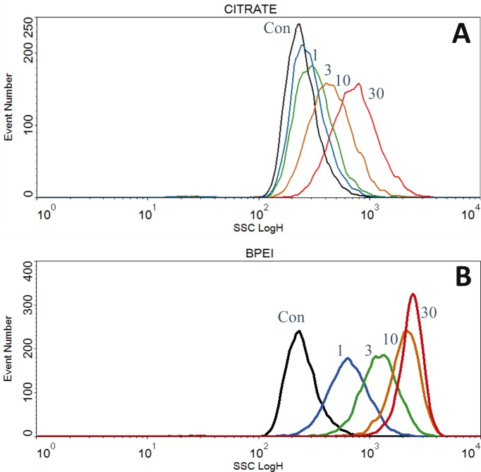
This examine in contrast the relative mobile uptake of 80 nm silver nanoparticles (AgNP) with four totally different coatings together with: branched polyethyleneimine (bPEI), citrate (CIT), polyvinylpyrrolidone (PVP), and polyethylene glycol (PEG).
A gold nanoparticle PVP was additionally in comparison with the silver nanoparticles. Biophysical parameters of mobile uptake and results included flow cytometry facet scatter (SSC) depth, nuclear mild scatter, cell cycle distributions, floor plasmonic resonance (SPR), fluorescence microscopy of mitochondrial gross construction, and darkfield hyperspectral imaging.
The AgNP-bPEI had been positively charged and entered cells at the next price than the negatively or neutrally charged particles. The AgNP-bPEI had been poisonous to the cells at decrease doses than the opposite coatings which resulted in mitochondria being reworked from a standard string-like look to small spherical beaded buildings.
Hyperspectral imaging confirmed that AgNP-bPEI and AgNP-CIT agglomerated within the cells and on the slides, which was evident by longer spectral wavelengths of scattered mild in comparison with AgNP-PEG and AgNP-PVP particles.
In unfixed cells, AgNP-CIT and AgNP-bPEI had increased SPR than both AgNP-PEG or AgNP-PVP particles, presumably on account of better intracellular agglomeration. After 24 hr. incubation with AgNP-bPEI, there was a dose-dependent lower within the G1 section and a rise within the G2/M and S phases of the cell cycle suggestive of cell cycle inhibition.
The nuclei of all of the AgNP handled cells confirmed a dose-dependent improve in nanoparticles following non-ionic detergent remedy during which the nuclei retained extra-nuclear AgNP, suggesting that nanoparticles had been hooked up to the nuclei or cytoplasm and not eliminated by detergent lysis. In abstract, positively charged AgNP-bPEI elevated particle mobile uptake.
Particles agglomerated within the peri-nuclear area, elevated mitochondrial toxicity, disturbed the cell cycle, and brought on irregular adherence of extranuclear materials to the nucleus after detergent lysis of cells. These outcomes illustrate the significance of nanoparticle floor coatings and cost in figuring out probably poisonous mobile interactions.
Quantitative section imaging of cells in a flow cytometry association using Michelson interferometer-based off-axis digital holographic microscopy.
We mixed Michelson-interferometer-based off-axis digital holographic microscopy (DHM) with a typical flow cytometry (FCM) association.
Utilizing object recognition procedures and holographic autofocusing in the course of the numerical reconstruction of the acquired off-axis holograms, sharply centered quantitative section photographs of suspended cells in flow had been retrieved with out labeling, from which biophysical mobile options of distinct cells, comparable to cell radius, refractive index and dry mass, might be subsequently retrieved in an automatic method. The efficiency of the proposed idea was first characterised by investigations on microspheres that had been utilized as check requirements.
Then, we analyzed two varieties of pancreatic tumor cells with totally different morphology to additional confirm the applicability of the proposed technique for quantitative dwell cell imaging.
The retrieved biophysical datasets from cells in flow are present in good settlement with outcomes from comparative investigations with beforehand developed DHM strategies beneath static situations, which demonstrates the effectiveness and reliability of our strategy.
Our outcomes contribute to the institution of DHM in imaging FCM and prospect to broaden the appliance spectrum of FCM by offering complementary quantitative imaging in addition to extra biophysical cell parameters which aren’t accessible in present high-throughput FCM measurements.